Axons in brain cells resemble a string of pearls rather than smooth tubes, according to Johns Hopkins researchers. This discovery, aided by advanced imaging and modeling, reveals how physical and membrane properties influence axon structure and function, challenging long-held beliefs and offering insights into brain signaling and disease.
Biology textbooks may require revision, according to Johns Hopkins Medicine scientists, who have presented new evidence suggesting that an armlike structure of mammalian brain cells might have a different shape than what scientists have assumed for over a century.
Their study on mouse brain cells shows that the cells’ axons — the armlike structures that reach out and exchange information with other brain cells — are not the cylindrical tubes often pictured in books and on websites but more like pearls on a string.
A report on the findings was recently published in the journal Nature Neuroscience.
“Understanding the structure of axons is important for understanding brain cell signaling,” says Shigeki Watanabe, Ph.D., associate professor of cell biology and neuroscience at the Johns Hopkins University School of Medicine. “Axons are the cables that connect our brain tissue, enabling learning, memory and other functions.”
Scientists have known that pearl-like structures in axons, referred to as axon beading, can develop in dying brain cells and in people with Parkinson’s and other neurodegenerative diseases due to the loss of membrane and skeletal integrity in neurons.
Under normal conditions, axons are thought to be shaped like tubes with a mostly constant diameter and occasional bubble-like structures (synaptic varicosities that hold globs of neurotransmitters, which enable signaling to other brain cells).
Investigating Axon Pearling
Watanabe had initially seen repeated axon pearling in the nervous system of worms and grew more curious about the structures after a discussion with Swiss scientist Graham Knott, Ph.D. A research team from Harvard University had published a study in 2012 that identified repeated “skeletal” components in axons, so the pair of researchers discussed experiments to get rid of the axon skeleton to see if the pearl structures disappear, says Watanabe.
Johns Hopkins graduate student and study first author Jacqueline Griswold tested the idea but found no effect on axon pearling.
Then, Watanabe and Griswold worked with theoretical biophysics colleague Padmini Rangamani, Ph.D., professor of pharmacology at the University of California San Diego School of Medicine, to look more closely at axons’ physical properties.
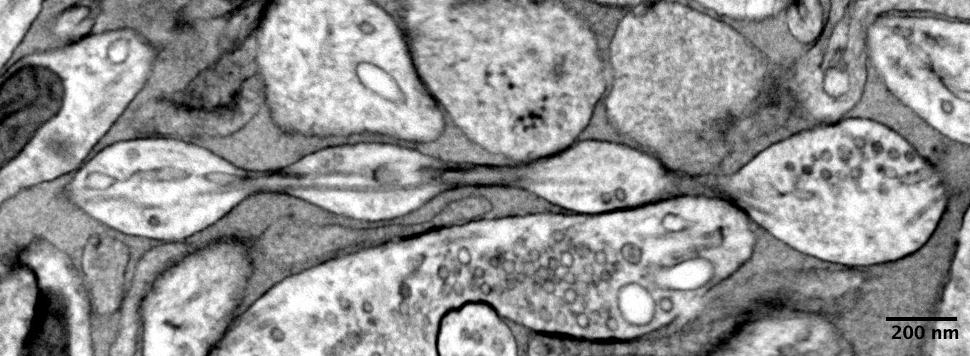
To be able to see axons on brain cells (neurons), which are 100 times smaller than the width of a human hair, the scientists used high pressure freezing electron microscopy. Like standard electron microscopy, which shoots beams of electrons at a cell to outline its structure, Watanabe and his team froze mouse neurons to preserve the structures’ shape.
“To see
The scientists named the pearl-like structures in which the axon swells “non-synaptic varicosities.”
“These findings challenge a century of understanding about axon structure,” says Watanabe.
Insights from Mathematical Modeling and Experiments
The scientists also used mathematical modeling to see if the axon membrane influenced the shape or presence of the pearl on a string structure. They found that simple mechanical models could be used to explain these structures very effectively.
Furthermore, experiments with the mathematical model and mouse brain samples showed that increasing the concentration of sugars in the solution around the axon or decreasing tension in the axonal membranes reduced the pearl structures’ size.
In another experiment, the scientists removed cholesterol from the neuron’s membrane to make it less stiff and more fluid-like. Under this condition, they found less pearling in both mathematical models and mouse neurons, along with reduced ability of the axon to transmit electrical signals.
“A wider space in the axons allows ions [chemical particles] to pass through more quickly and avoid traffic jams,” says Watanabe.
The scientists also applied high frequency electrical stimulation to the mouse neurons, which made pearled structures along axons swell, on average, 8% longer and 17% wider for at least 30 min after stimulation and increased the speed of electrical signals. However, when cholesterol was removed from the membrane, the axon’s pearls lost their swollen state and had no change in the speed of electrical signals.
The research team plans to examine axonal “arms” in human brain tissue taken with permission from people having brain surgery and those who have died from neurodegenerative diseases. This work formed the basis of a recently awarded Multiple Principal Investigator grant to Watanabe and Rangamani from the National Institute of Mental Health.
Reference: “Membrane mechanics dictate axonal pearls-on-a-string morphology and function” by Jacqueline M. Griswold, Mayte Bonilla-Quintana, Renee Pepper, Christopher T. Lee, Sumana Raychaudhuri, Siyi Ma, Quan Gan, Sarah Syed, Cuncheng Zhu, Miriam Bell, Mitsuo Suga, Yuuki Yamaguchi, Ronan Chéreau, U. Valentin Nägerl, Graham Knott, Padmini Rangamani and Shigeki Watanabe, 2 December 2024, Nature Neuroscience.
DOI: 10.1038/s41593-024-01813-1
Funds for the research were provided by the Johns Hopkins University School of Medicine, the Marine Biological Laboratory Whitman Fellowship, the Chan Zuckerberg Initiative Collaborative Pair Grant and Supplement Award, the Brain Research Foundation Scientific Innovations Award, a Helis Foundation award, the